 I. F. Anderson Farms
 Science and Technology
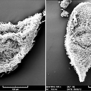 Opalinids
501-918-3025
calsfoundation@cals.org
calsfoundation@cals.org
Opalinids are a small group of peculiar cosmopolitan organisms that belong to the kingdom Protista. Recent classification places the opalinids as heterotrophic stramenopiles (heterokonts) within the phylum Placidozoa, class Opalinea, and order Slopalinida. There are over 200 species, and, although opalinids are typically endocommensals (that is, living within the host without affecting it) in the large intestine and cloaca of anurans (frogs and toads), they have also been reported from fish, salamanders, reptiles, and some invertebrates (mollusks and insects). They are of no medical or economic importance, but they are interesting because their reproductive cycles are apparently controlled by the host’s hormones. In addition, opalinids are routinely encountered in dissections of frogs in college biology laboratories, and most students are surprised to know that they even exist. Several surveys have reported opalinids from Arkansas anurans, including from a toad in Lonoke County and chorus frogs in Union County. The state supports twenty-nine species and subspecies of anurans, so it is likely that a number of species of opalinids occur in frogs and toads in the state.
Initially, biologists thought that the hair-like structures that covered opalinids’ surface were cilia, and they placed them within the phylum Ciliophora (ciliates). However, in the early twentieth century, other aspects of their biology clearly helped differentiate them from the ciliates, and they were placed in the phylum Sarcomastigophora, with the amoebae and flagellates. In the 1980s, detailed ultrastructural studies of Opalina ranarum using electron microscopy revealed that they share morphologies with the heterokonts of the family Proteromonadidae. Then, in 1985, a new order, the Slopalinida was proposed to include the members of the families Proteromonadidae and Opalinidae. In 2004, the first reliable opaline molecular sequence data supported the monophyletic origin of the order Slopalinida. Therefore, researchers now consider the opalines to be a family (Opalinidae) within the order Slopalinida.
The cell surface of opalinids possesses numerous rows of flagella (kineties) in oblique and delicate parallel rows that allow interference of reflected light and opalescence; superficially, they look like ciliates. For traditional (typical) opalinids, the anterior end of the organism is defined as the direction of travel. The anterior margin is usually hyaline, against which the anterior ends of the kineties abut, which is termed the falx. They have a left-handed spiral path of movement, no mouth, no contractile vacuole, but large nuclei in binucleate forms and small in multinucleate forms; there is no macro- or micronucleus.
Lacking a mouth, opalinids get nutrition via pinocytosis (ingestion of liquid into a cell by using small vesicles from the membrane) and reproduce by means of syngamy (sexual reproduction) with union or fusion of two gametes, which differ in size and/or form (anisogamy). While some experts consider the opalines to be parasites, evidence suggests they are actually endocommensals that do not injure their hosts. Since the host absorbs nutrients from what it ingests in the small intestine, the opalines are likely not robbing them of essential nutrients. It is therefore assumed that the opalines are simply living off the leftover nutrients in the feces, possibly supplemented by the biochemical contributions of the lush bacterial flora that also reside there. Anurans often contain many hundreds and sometimes thousands of opalines in their lower bowel and feces, and they appear to be completely healthy, with no evident pathologies in their intestinal or cloacal epithelia.
Very little is known about the life cycles of opalines in fish, reptilian, or arthropod hosts. However, the life cycle of a standard opalinid (like Opalina ranarum) includes an asexual stage of multinucleate trophozoites (trophonts) in the cloacal area of adult frogs. They divide by binary fission most of year and yield more trophonts, and from late winter to early spring, production of cysts and the sexual reproductive cycle occurs. About two weeks prior to frogs arriving at breeding sites, hastening of binary fission occurs. Numerous smaller forms (tomonts), about thirty to ninety micrometers long and with fewer nuclei, are generated (referred to as palintony). These encyst, and cysts thirty to seventy micrometers in diameter and possessing two to twelve nuclei each are passed out into the aquatic environment with the frog feces. When these are consumed by tadpoles, the gamonts excyst, and numerous divisions, including meiosis, occur. Two types of gametes are produced: larger (macrogametes) and smaller (microgametes). These anisogamous gametes fuse (via syngamy), and the zygote divides often and encysts as a zygocyst or cytozygote. Cysts then exit with feces, are eaten by new tadpoles, and excyst. They grow and become multinucleate and reside in the anuran gut until the following breeding season.
There are two families: the Proteromonadidae and Opalinidae. The former family includes Proteromonas (four species), with two apical flagella, a single nucleus, a large dictyosomal (Golgi body) complex located near the anterior end, and a surface that is not markedly ridged. They have been reported from salamanders, lizards, and mammals. Another in this family is Karotomorpha (several species) with four long apical flagella, a single nucleus, a large dictyosomal complex located near the anterior end, and a surface that is markedly ridged. This genus has been reported from amphibians, particularly newts (Taricha sp.) from the western United States.
The Opalinidae family includes five recognized genera as follows: Cepedea, Opalina, Protoopalina, Protozelleriella, and Zelleriella. These genera differ morphologically in terms of the number of nuclei, the appearance and location of the falx (two short, sickle-shaped rows of flagella), and whether the long rows of flagella (kineties) cover the body evenly or if there is a “bald spot.” Due to the differences in body shape among the different life cycle stages within a species, whether overall body shape (flat or cylindrical) can be used to differentiate the genera has not yet been determined.
The Cepedea are known from frogs and toads. They are multinucleate, with a short, broad, axial falx almost parallel to the anteroposterior axis of the cell; kineties cover the body evenly; and most species have an elongate, cylindroid body. Opalina spp., from various amphibians, are multinucleate, with a long, thin, marginal falx almost perpendicular to the anterior posterior axis of the cell; kineties cover the body evenly (no hyaline margin devoid of flagella); there are broad and elongate forms with a dorsoventral suture line at the posterior of the cell; and most species are broadly ellipsoidal or ovoidal and dorso-ventrally flattened. Protoopalina spp. have been reported exclusively from frogs and toads and are binucleate, with a short, broad, axial falx almost parallel to the anteroposterior axis of the cell and kineties covering the body evenly, and most species have an elongate, cylindroid body. Protozelleriella spp. are known from toads and are binucleate, with a long, thin, marginal falx almost perpendicular to the anterior posterior axis of the cell. The peripheral hyaline margin is devoid of flagella; there is a crenulate posterior margin and a mushroom-shaped profile. The Zelleriella have been reported from amphibians but rarely reptiles, and are binucleate with a long, thin, marginal falx almost perpendicular to the anterior posterior axis of the cell; most species are broadly ellipsoidal to ovoidal and dorsoventrally flattened, and a few species possess a mushroom-shaped body.
There are only a few reports of opalines in fishes, and even fewer documenting opalines from reptilian or salamander hosts. Their rarity outside of anuran hosts had led many to speculate that the other hosts represent accidental infections—for example, a snake eating a frog infected with opalinids. However, opalines have been found in marine fishes that are restricted to freshwater environments that have no access to anurans. In addition, populations of opalines in fish hosts are often very high, suggesting that they are probably reproducing. In amphibians other than anurans, Protoopalina mitotica has been reported from salamanders. In reptiles, Opalina spp. have been reported from monitor lizards (Varanus spp.), Protoopalina sp. has been documented from a Nile monitor, Varanus niloticus from Africa, Zelleriella boipevae has been reported from the false yarara, Xenodon merremii from South America, Z. jaegeri has been found in a Jaeger’s ground snake, Erythrolamprus jaegeri from South America, and an unknown opalinid was reported from a tortoise, Testudo sp.
Several surveys have reported opalinids from Arkansas anurans. A new species of Protoopalina was described in 1995 from an eastern narrowmouth toad (Gastrophryne carolinensis) collected from Anderson’s Minnow Farm in Lonoke County. Opalina spp. have been documented from Cajun chorus frogs (Pseudacris fouquettei) from near El Dorado (Union County), from bird-voiced treefrogs (Hyla avivoca) from Conway and Lafayette counties, and from pickerel frogs (Rana palustris) from an abandoned mine in Independence County. Since Arkansas supports twenty-nine species and subspecies of anurans, other frogs and toads in the state likely harbor additional species of opalinids.
For additional information:
Barnard, Susan M., and Steve J. Upton. A Veterinary Guide to the Parasites of Reptiles. Vol. 1. Malabar, FL: Krieger Publishing Company, 1994.
Corliss, John O. “The Opalinid Infusorians: Flagellates or Ciliates?” Journal of Protozoology 2 (1955): 107–114.
Delvinquier, Ben L. J., and Sherwin S. Desser. “Opalinidae (Sarcomastigophora) in North American Amphibia.” Systematic Parasitology 33 (1996): 33–51.
Delvinquier, Ben L. J., Sherwin S. Desser, and Jerry Johnson. “Opalinidae (Sarcomastigophora) in North American Amphibia. Genus Protoopalina Metcalf, 1918.” Systematic Parasitology 32 (1995): 141–147.
Delvinquier, Ben L. J. and David J. Patterson. The Opalinids. In Parasitic Protozoa, 2nd ed. Vol. 3. Edited by J. P. Kreier and J. R. Baker. San Diego: Academic Press, 1992.
———. Order Slopalinida Patterson, 1989. In Guide to the Protozoa, 2nd ed. Lawrence, KS: Allen Press, 2002.
Frank, W. Non-Hemoparasitic Protozoans. In Diseases of Amphibians and Reptiles, edited by G. L. Hoff, Fredric L. Frye, and Elliot R. Jacobson. New York: Plenum Press, 1984.
Hoevers, J. D., and K. F. Snowden. “Analysis of the ITS Region and Partial SSU and LSU rRNA Genes of Blastocystis and Proteromonas lacertae .” Parasitology 131 (2005): 187–196.
McAllister, Chris T., Charles R. Bursey, Matthew B. Connior, and Stanley E. Trauth. “Symbiotic Protozoa and Helminth Parasites of the Cajun Chorus Frog, Pseudacris fouquettei (Anura: Hylidae), from Southern Arkansas and Northeastern Texas.” Comparative Parasitology 80 (2013): 96–104.
McAllister, Chris T., Charles R. Bursey, and Stanley E. Trauth. “Parasites of the Pickerel Frog, Rana palustris (Anura: Ranidae) from the Southern Part of its Range.” Southwestern Naturalist 40 (1995): 111–116.
McAllister, Chris T., Stanley E. Trauth, Steve J. Upton, and David H. Jamieson. “Endoparasites of the Bird-Voiced Treefrog, Hyla avivoca (Anura: Hylidae), from Arkansas.” Journal of the Helminthological Society of Washington 60 (1993): 140–143.
Metcalf, M. M. “The Bell-Toads and their Opalinid Parasites.” American Naturalist 62 (1928): 5–21.
———. “The Classification of the Opalinidae.” Science 52 (1920): 135–136.
———. “Further Studies on the Opalinid Ciliate Infusorians and Their Hosts.” Proceedings of the United States National Museum 87 (1940): 465–634.
———. “Opalina and the Origin of the Ciliata.” Anatomical Record 14 (1918): 88–89.
———. “The Opalinid Ciliate Infusorians.” Bulletin of the United States National Museum 120 (1923): 1–484.
Patterson, David J. “The Fine Structure of Opalina ranarum (Family Opalinidae): Opalinid Phylogeny and Classification.” Protistologica 21(1985): 413–428.
Roberts, Larry S., John Janovy Jr., and Steven Nadler. Foundations of Parasitology. New York: McGraw-Hill Higher Education, 2012.
Wessenberg, H. “Studies on the Life Cycle and Morphogenesis of Opalina.” University of California Studies in Zoology 61 (1961): 315–370.
Chris T. McAllister
Eastern Oklahoma State College


Comments
No comments on this entry yet.