
501-918-3025
calsfoundation@cals.org
calsfoundation@cals.org
Slime molds are among the more interesting yet relatively little known organisms found in Arkansas. They do not have a particularly attractive name, but some examples produce fruiting bodies that are miniature objects of considerable beauty. The organisms commonly referred to as slime molds actually belong to two different taxonomic groups—the myxomycetes and the dictyostelids. Members of both groups are common-to-abundant organisms in forests throughout Arkansas, but only the myxomycetes (also called plasmodial slime molds) can be observed directly in nature. As a result, the myxomycetes are by far the better known group, and to most people (including many biologists), they are the only “slime molds” of which they are aware. However, the dictyostelids (also known as cellular slime molds) are often exceedingly abundant in the microhabitats where they occur.
As used here, the term “microhabitat” refers to some portion of a larger habitat. For example, a forest as a habitat is composed of a number of smaller microhabitats, including the microhabitat represented by the logs, stumps, and other pieces of woody debris found on the forest floor. Other microhabitats are the layer of dead leaves, also found on the forest floor, the forest soil, and the bark surface of living trees. At least some slime molds are associated with all of these microhabitats, but the dictyostelids have a more limited distribution than do the myxomycetes. Dictyostelids are most commonly associated with the microhabitat represented by the surface humus layers of forest soil, whereas the ecological distribution of myxomycetes encompasses all of the different microhabitats mentioned above.
Dictyostelids
The dictyostelids are a relatively homogeneous group of approximately 150 described species, about twenty of which are known to occur in Arkansas. The usual method for studying dictyostelids involves collecting small samples (usually no more than half an ounce or so) of soil in the field, returning these samples to a laboratory, diluting and suspending a portion of each sample in distilled water, and then adding this to the surface of a weak nutrient agar in a Petri dish along with a bacterium such as E. coli to serve as a food source for the dictyostelid amoebae present. The dish is incubated at room temperature for two or three days and then checked for the presence of the fruiting bodies of dictyostelids with the use of a stereomicroscope. More often than not, cultures prepared in this manner will yield the fruiting bodies of one or more species of dictyostelids.
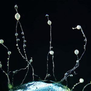
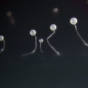
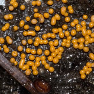
The fruiting body of a dictyostelid consists of a slender stalk bearing one or more clusters (called sori) of spores. In some species, there is only a single cluster of spores at the apex of the stalk, but in other species the main stalk gives rise to one or more branches, with each of these bearing a cluster of spores. The stalk is essentially colorless, and the same is often the case for the cluster of spores. However, in a few species, the cluster of spores is pigmented, with yellow and violet the most common. Although fruiting bodies can exceed a height of more than a millimeter, they are not always obvious to someone who has no previous experience working with dictyostelids. The reason for this is that the fruiting bodies of dictyostelids closely resemble the spore-producing structures made by some of the microfungi that characteristically appear in cultures prepared with samples of soil. However, the fruiting body of a dictyostelid has a fundamentally different origin than any of the structures produced by soil microfungi. For most of their life cycle, dictyostelids exist as uninucleate amoeboid cells. These feed upon soil bacteria and undergo cell division to produce new cells. Where bacteria are abundant, which is usually the case in the upper humus layers where dictyostelids are most common, repeated cell divisions result in large populations of their amoeboid cells. However, when their food supply of bacteria becomes depleted, the cells do something rather remarkable. They migrate in streams to what is known as an aggregation center, which is nothing more than a large mass of individual cells. This process, which is coordinated by the production of special biochemical attractants, ultimately results in the formation of one or more elongated slug-shaped structures (referred to as “slugs” or “pseudoplasmodia”) in which each of the original cells maintains its individual integrity. Pseudoplasmodia may migrate for short distances in some species or transform directly into a fruiting body, with some of the original cells giving rise to spores and others to the stalk. Aggregation and fruiting body formation represent an asexual dispersal process more than actual reproduction, but sexual reproduction is known for many species of dictyostelids. Interestingly, because the aggregation of amoeboid cells to form a pseudoplasmodium is very similar to what must have happened over the course of evolution when unicellular organisms gave rise to the first multicellular forms, dictyostelids have been the subject of considerable research in the areas of cell and molecular biology.
Although most commonly thought of as inhabitants of forest soils, dictyostelids also occur in some abundance in soils from other types of habitats, including agricultural fields. Among the most frequently encountered species in Arkansas are Dictyostelium minutum, Dictyostelium purpureum, Polysphondylium pallidum, and Polysphondylium violaceum. Dictyostelids are often surprisingly common in caves, and many of the Ozark caves investigated to date have yielded records of these organisms. Two of the species of dictyostelids known from Arkansas—Dictyostelium caveatum and Dictyostelium rosarium—have been found only in caves.
Myxomycetes
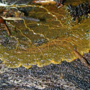
The life cycle of a myxomycete contains two very different trophic (or feeding) stages. The first of these consists of uninucleate amoeboid cells (which may or may not be flagellated), and the second is represented by a distinctive multinucleate structure referred to as a plasmodium (the basis for the common name “plasmodial slime molds”). The plasmodium is essentially a large amoeboid cell that can move about (albeit rather slowly), feeding upon bacteria, other microscopic organisms, fungal spores, and tiny bits of organic matter. Plasmodia can reach a size of several centimeters, with truly extraordinary examples sometimes exceeding a meter across. They are not difficult to find in nature during the summer and fall. The best way to look for them is to pull away pieces of loose bark from a moist decaying log or stump.
Under favorable conditions, the plasmodium ultimately gives rise to one or more fruiting bodies containing spores. The spores are apparently largely wind-dispersed, and if they land in a suitable place, germination occurs to produce the uninucleate amoeboid cell. These divide by simple cell division to build up large populations in the microhabitats where they occur. Two of these uninucleate amoeboid cells, which are haploid (possessing a single set of chromosomes), fuse to form a diploid (possessing two sets of chromosomes) zygote, which then develops into the plasmodium, in which all of the nuclei present are diploid. Under appropriate conditions, such as drying out of the microhabitat, a plasmodium gives rise to one or more fruiting bodies, within which meiosis (reduction division) occurs when the spores are produced.
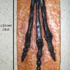
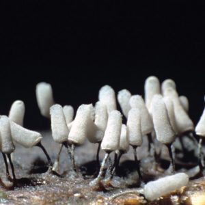
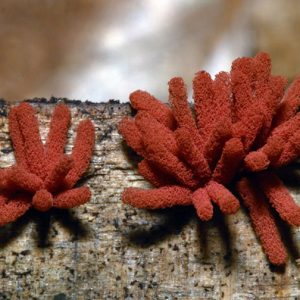
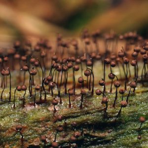
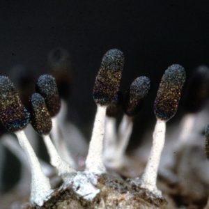
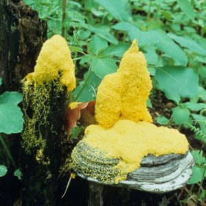
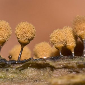
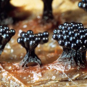
The fruiting bodies produced by myxomycetes are often no more than an eighth of an inch in height, but a single relatively large fruiting may consist of several hundred individual fruiting bodies and extend over an area of several square inches. The fruiting bodies of some myxomycetes resemble tiny mushrooms, whereas others have shapes that are more difficult to characterize. Although they are easily overlooked in nature, careful examination of decaying logs and stumps during late summer and early fall, especially after a period of rainy weather, will almost invariably turn up the fruiting bodies of several different species. Some of the more commonly encountered myxomycetes in the forests of Arkansas are Lycogala epidendrum, with fruiting bodies that look like miniature puffballs; Trichia favoginea, in which the yellow, egg-shaped fruiting bodies occur in dense clusters; Arcyria denudata, characterized by bright red, elongated fruiting bodies that appear to have the general texture of cotton candy; and Stemonitis fusca, with dark brown to almost black, hair-like fruiting bodies that occur in little tufts on decaying wood. Because of their size, the fruiting bodies of most myxomycetes are best appreciated when viewed with a magnifying glass or a hand lens. However, this is not always the case. The largest fruiting bodies of any myxomycete known from Arkansas are produced by Fuligo septica, which is often common on the beds of mulch found around ornamental shrubs. Fruiting bodies in this species sometimes reach the size of a dinner plate.
Unlike many other groups of organisms found in Arkansas, the myxomycetes present in a particular locality in the state are likely to be the very same species present in any other locality. This same situation is generally true for the entire temperate zone of the world. However, a few species of myxomycetes do appear to be confined largely to such special habitats as tropical rainforests and deserts.
For additional information:
Eliasson, Uno H., Harold W. Keller, and James A. Hutchison. “Myxomycetes from Arkansas.” Mycotaxon 32 (1988): 375–398.
Landolt, John C., Steven L. Stephenson, and Adam W. Rollins. “Dictyostelid Cellular Slime Molds of Arkansas.” Castanea 74 (2009): 353–359.
Landolt, John C., Steven L. Stephenson, and Michael E. Slay. “Dictyostelid Cellular Slime Molds from Caves.” Cave and Karst Journal 68 (2006): 22–26.
Raper, Kenneth B. The Dictyostelids. Princeton, NJ: Princeton University Press, 1984.
Stephenson, Steven L., and Carlos Alvarado, eds. Myxomycetes: Biology, Systematics, Biogeography, and Ecology. Cambridge, MA: Academic Press, 2017.
Stephenson, Steven L., and Henry Stempen. Myxomycetes: A Handbook of Slime Molds. Portland, OR: Timber Press, 1994.
Trimble, Courtney. “Spore Dispersal of Slime Molds and Higher Fungi via Animal Vectors.” MS thesis, University of Arkansas, 2021. Online at https://scholarworks.uark.edu/etd/4085/ (accessed July 6, 2022).
Steven L. Stephenson
University of Arkansas, Fayetteville


Comments
No comments on this entry yet.